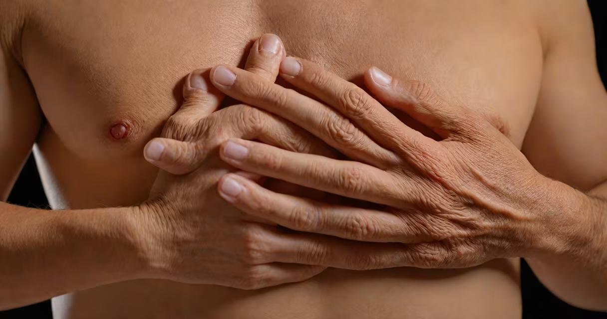
Costochondritis is inflammation of the cartilage connecting the ribs to the sternum, causing sharp, localized chest pain often mistaken for heart issues. It affects people of all ages but is most common in adults between 20 and 50, with women more frequently diagnosed than men.
This condition typically develops after physical strain, viral infection, or repetitive upper-body movements. Pain worsens with deep breathing, coughing, or pressure on the chest wall. Unlike cardiac events, costochondritis pain doesn't radiate to the arm or jaw and usually stays confined to one side of the chest.
According to the American College of Rheumatology, costochondritis accounts for approximately 10-20% of non-traumatic chest pain cases seen in primary care settings. It's rarely life-threatening but can disrupt daily activities due to persistent discomfort.
The inflammation occurs in the costosternal joints-the junctions where the upper rib bones meet the breastbone via cartilage. When irritated, these tissues swell and become tender, triggering pain with movement or touch.
Pain onset is often sudden and may follow a recent cold, heavy lifting, or prolonged sitting with poor posture. The sensation is sharp and localized, unlike the dull, crushing ache associated with heart attacks. Patients often describe it as "stabbing" or "aching," and it typically lasts days to weeks, though chronic cases can persist for months.
A key distinguishing feature is tenderness upon palpation. A doctor presses gently along the rib-sternum junctions; if pain increases at specific points, it supports a diagnosis of costochondritis. Imaging like X-rays or CT scans usually shows no abnormalities, which helps rule out structural or cardiac causes.
While costochondritis is a single clinical entity, its onset can be linked to different underlying factors. These fall into four categories:
| Trigger Type | Common Causes | Typical Presentation |
|---|---|---|
| Postural/Repetitive Strain | Heavy lifting, poor ergonomics, repetitive motions | Pain on one side, worsened by reaching or twisting |
| Viral Infection | Upper respiratory infections, post-viral syndrome | Acute onset, often bilateral, resolves within 6 weeks |
| Trauma | Blunt chest impact, falls, car accidents | Localized tenderness, swelling, bruising |
| Autoimmune or Systemic Conditions | Ankylosing spondylitis, lupus, rheumatoid arthritis | Chronic pain, multiple joint involvement, positive blood markers |
When no clear cause is identified, it's termed idiopathic costochondritis. This form accounts for about 60% of cases reported in the Journal of Clinical Rheumatology.
Healthcare providers use costochondritis patient info to differentiate it from serious conditions like myocardial infarction, pulmonary embolism, or pleurisy. Misdiagnosis leads to unnecessary testing and anxiety.
For example, a 34-year-old woman presented with left-sided chest pain lasting three days. She had no risk factors for heart disease-no smoking, normal cholesterol, and no family history. On exam, she winced when her left sixth rib was pressed. An EKG showed no ischemia. After ruling out cardiac causes, the diagnosis of costochondritis was confirmed. She improved with NSAIDs and rest within two weeks.
Diagnostic criteria from the European League Against Rheumatism emphasize tenderness at the costosternal junctions and absence of systemic signs like fever or weight loss. If these are absent, other diagnoses such as Tietze's syndrome (which includes swelling) or referred pain from the spine or abdomen must be considered.
Many patients assume costochondritis is a sign of heart disease. This is incorrect. The pain location-center or slightly left of center, below the clavicles-is distinct from cardiac pain, which often radiates to the neck, jaw, or left arm.
Another misconception is that it requires imaging. Most cases don't. A 2021 study in BMJ Open found that 87% of costochondritis cases were correctly diagnosed based on history and physical exam alone, without needing X-rays or MRIs.
Some believe it's always short-lived. While most resolve in weeks, chronic cases do occur. A 2019 retrospective review in Rheumatology International found that 12% of patients experienced symptoms beyond six months, especially those with comorbid fibromyalgia or anxiety disorders.
First-line treatment involves nonsteroidal anti-inflammatory drugs (NSAIDs) like ibuprofen 600 mg twice daily for up to seven days. Topical lidocaine patches applied directly over painful areas have shown moderate efficacy in reducing localized pain, according to a 2020 double-blind trial published in Pain Medicine.
Physical therapy techniques such as diaphragmatic breathing exercises and postural correction reduce muscle tension around the chest wall. A 2022 pilot study found that patients who practiced daily breathing drills reported 40% less pain intensity after four weeks compared to controls.
Rest from aggravating activities is critical. Avoid pushing heavy objects or doing push-ups during flare-ups. Sleep position matters too-lying on the affected side can increase pressure. Sleeping on the back with a pillow under the upper back reduces strain.
Seek immediate care if chest pain is new, severe, or accompanied by shortness of breath, dizziness, sweating, or nausea. These symptoms suggest a cardiac event and require urgent evaluation.
Also consult a provider if pain persists beyond six weeks despite home management, spreads to the shoulder or back, or wakes you at night. These changes may indicate another condition such as cervical radiculopathy or esophageal reflux.
For most patients, costochondritis is manageable with conservative care. Understanding the nature of the pain and knowing when to act prevents unnecessary worry and medical visits.
Costochondritis patient info clarifies that while uncomfortable, this condition is not dangerous. With accurate diagnosis and targeted relief methods, recovery is typical within weeks. Awareness of red flags ensures timely intervention when needed.