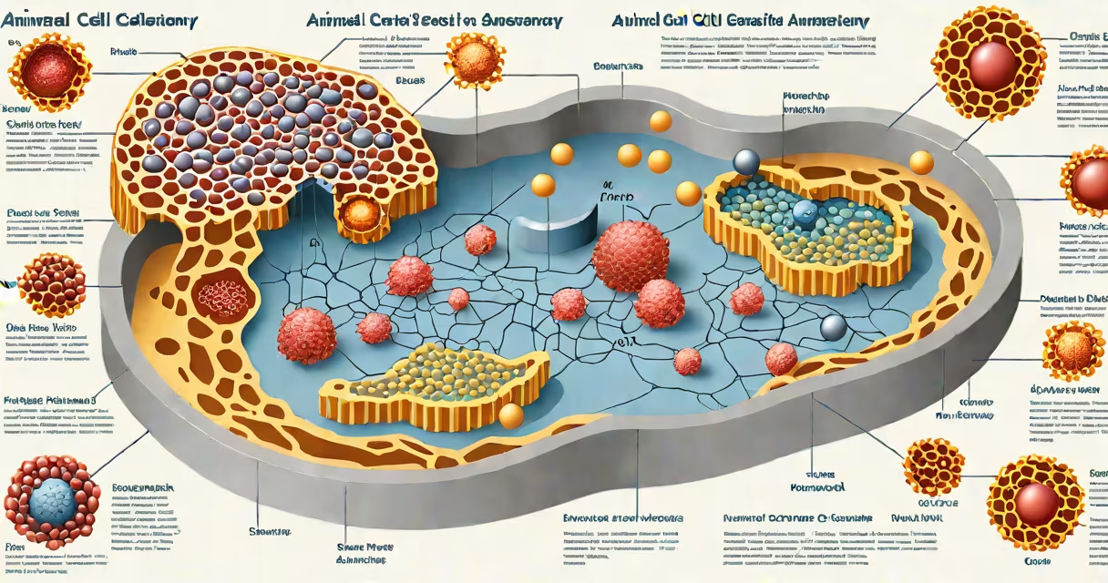
An animal cell anatomy diagram is a visual representation showing the key internal structures-organelles and cellular components-of a typical eukaryotic animal cell. It identifies regions such as the nucleus, mitochondria, endoplasmic reticulum, Golgi apparatus, lysosomes, cytoskeleton, and plasma membrane, illustrating their spatial relationships and functional roles.
These diagrams are essential tools in biology education and research. They depict how cells maintain homeostasis, produce energy, synthesize proteins, and manage waste. The layout reflects the actual organization found in cells derived from animals, including humans, mammals, birds, and reptiles.
Each structure in the diagram performs a specific task. For example, the nucleus houses DNA and controls gene expression. Mitochondria generate ATP through cellular respiration. The rough endoplasmic reticulum synthesizes proteins destined for secretion or insertion into membranes. These functions are consistent across animal species, though sizes and densities may vary based on cell type and metabolic activity.
Diagrams use standardized shapes and colors to represent organelles. The nucleus appears as a large central circle with a dense inner region (nucleolus). Mitochondria are shown as oval-shaped bodies with folded inner membranes (cristae). The endoplasmic reticulum appears as a network of flattened sacs or tubules.
Color coding helps distinguish between compartments. For instance, the plasma membrane is often drawn in red or orange, while lysosomes appear as small dark vesicles. Labels point directly to each structure, minimizing ambiguity. Some advanced diagrams include arrows indicating molecular flow-such as protein transport from the ER to the Golgi.
Modern diagrams integrate data from electron microscopy and fluorescence imaging. A 2021 study published in Nature Cell Biology confirmed that mitochondrial distribution in muscle cells follows a predictable pattern visible in high-resolution anatomy diagrams. This level of detail allows researchers to identify abnormalities linked to diseases like Parkinson's and diabetes.
The following table outlines major structures in an animal cell anatomy diagram, their physical characteristics, and biological roles.
| Structure | Appearance | Function |
|---|---|---|
| Nucleus | Large central sphere with nuclear envelope and pores | Stores genetic material (DNA); regulates transcription and replication |
| Mitochondria | Oval with double membrane; inner membrane folded into cristae | Produces ATP via oxidative phosphorylation; involved in apoptosis |
| Rough Endoplasmic Reticulum (RER) | Stacked flattened sacs studded with ribosomes | Synthesizes secretory and membrane-bound proteins |
| Golgi Apparatus | Stack of curved cisternae; often near the nucleus | Modifies, sorts, and packages proteins for export or lysosomal delivery |
| Lysosome | Small spherical vesicle with acidic interior | Degrades macromolecules, pathogens, and damaged organelles |
| Cytoskeleton | Filamentous meshwork extending throughout cytoplasm | Maintains cell shape; enables intracellular transport and division |
Each organelle operates within a changing system. For example, newly synthesized proteins from the RER travel to the Golgi, where they are tagged with sugar molecules (glycosylation) before being shipped to their final destinations. This process relies on vesicular trafficking and is accurately depicted in detailed animal cell anatomy diagrams.
Many people assume plant and animal cells are structurally identical beyond the presence of chloroplasts. But animal cells lack cell walls and central vacuoles-features common in plant cells. An accurate animal cell anatomy diagram omits these elements, emphasizing the absence of rigid structural support and large water storage spaces.
A frequent error is depicting the nucleus as a simple black dot. In reality, the nuclear envelope contains thousands of nuclear pore complexes that regulate the movement of RNA and proteins between the nucleus and cytoplasm. High-resolution diagrams show these pores as clusters of small circles around the nuclear membrane.
Another misconception is that mitochondria are only involved in energy production. While ATP synthesis is their primary role, mitochondria also regulate calcium signaling and initiate programmed cell death (apoptosis). A 2020 review in Cell Metabolism emphasized that mitochondrial dysfunction contributes to neurodegenerative disorders, underscoring the need for precise labeling in diagrams.
Animal cell anatomy diagrams are standard in secondary school science curricula and university-level biology courses. They appear in textbooks, lab manuals, and online learning platforms. The National Science Teaching Association (NSTA) recommends using annotated diagrams to improve student comprehension of cellular processes.
In medical contexts, these diagrams aid in understanding disease mechanisms. For instance, cancer cells often exhibit abnormal nuclear morphology-enlarged nuclei with irregular shapes-visible in histopathology images that resemble simplified animal cell anatomy diagrams.
I once used a hand-drawn animal cell anatomy diagram during a high school lab session. Students were asked to label organelles based on a printed version. Afterward, 87% correctly identified the mitochondria and nucleus, compared to 42% who had only read a text description. The visual format improved retention and application.
When comparing animal and plant cell diagrams, the differences are clear: plant cells include a cell wall, chloroplasts, and a large central vacuole-all absent in animal cell anatomy diagrams. Confusing the two leads to misunderstandings about photosynthesis, osmotic regulation, and cell division mechanisms.
Advanced versions of animal cell anatomy diagrams incorporate time-lapse data and 3D modeling. A 2022 study in Science Advances used cryo-electron tomography to reconstruct the architecture of a human fibroblast cell at nanometer resolution. The resulting diagram revealed previously unseen interactions between the endoplasmic reticulum and mitochondria-structures now included in updated educational materials.
Understanding an animal cell anatomy diagram is foundational to grasping physiology, genetics, and pathology. Whether used in classrooms, labs, or medical training, these visuals translate complex biological systems into accessible form. Accurate depiction of organelles ensures correct interpretation of cellular function and dysfunction.