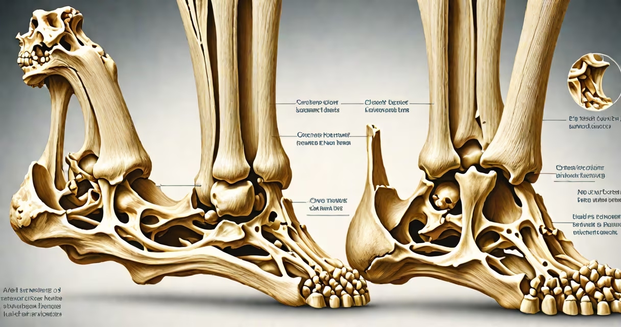
The ankle is a synovial hinge joint formed by the distal tibia and fibula articulating with the superior surface of the talus bone. This joint allows controlled motion primarily in one plane-dorsiflexion and plantarflexion-essential for standing, walking, and running.
As a hinge joint, the ankle restricts movement to flexion and extension. The tibia forms the medial and posterior walls of the joint, while the fibula contributes the lateral wall. Together, they create a mortise-like socket that locks the talus in place during weight-bearing activities. This structure provides stability and limits side-to-side motion, reducing injury risk during changing tasks.
According to Gray's Anatomy (41st edition), the ankle joint is classified as a ginglymus-a type of synovial joint that permits uniaxial movement. The joint capsule is reinforced by ligaments including the deltoid ligament medially and the lateral ligament complex (anterior talofibular, calcaneofibular, and posterior talofibular) laterally. These structures prevent excessive inversion and eversion, which could otherwise lead to sprains.
Motion at the ankle occurs when muscles in the lower leg contract. The tibialis anterior pulls the foot upward (dorsiflexion), while the gastrocnemius and soleus muscles pull the foot downward (plantarflexion). These movements are critical during the gait cycle: dorsiflexion occurs during heel strike to allow the foot to clear the ground, and plantarflexion powers push-off during walking and running.
A single step involves approximately 15 degrees of dorsiflexion and 45 degrees of plantarflexion. During sprinting, plantarflexion can reach up to 60 degrees. These ranges are limited by the bony architecture and soft tissue constraints. For example, the medial malleolus of the tibia prevents excessive eversion, while the lateral malleolus of the fibula limits inversion beyond 20 degrees.
When the foot lands, the ankle absorbs impact forces. Studies show that during running, the ankle experiences peak loads of up to 2.5 times body weight. The joint's ability to distribute force through its cartilage and synovial fluid reduces wear on bone surfaces and maintains long-term function.
The ankle includes two distinct joints: the true ankle joint (talocrural joint) and the subtalar joint. The talocrural joint is the primary hinge joint responsible for dorsiflexion and plantarflexion. The subtalar joint lies beneath the talus and connects the talus to the calcaneus. It enables inversion and eversion, allowing the foot to adapt to uneven terrain.
| Joint Type | Location | Movement | Ligament Support |
|---|---|---|---|
| Talocrural (true ankle) | Tibia-fibula & talus | Dorsiflexion, plantarflexion | Deltoid (medial), lateral ligament complex |
| Subtalar | Talus & calcaneus | Inversion, eversion | Calcaneofibular, cervical, interosseous ligaments |
| Tibiofibular syndesmosis | Proximal tibia & fibula | Minimal motion; stabilizes ankle | Interosseous membrane, anterior/posterior tibiofibular ligaments |
Many people confuse the ankle joint with the subtalar joint. The subtalar joint is often involved in chronic ankle instability after sprains. When the lateral ligaments are injured, the subtalar joint may compensate by increasing pronation, leading to altered gait patterns and secondary pain in the arch or knee.
One non-obvious fact: the ankle joint is not purely a hinge. While it functions as a uniaxial joint, subtle rotational motions occur due to slight asymmetry in the talus's articular surface. This allows micro-movements during standing that improve balance and reduce fatigue in the lower limb.
Some assume the ankle is a simple hinge, but it integrates multiple joint systems. A sprain involving the lateral ligaments affects both the talocrural and subtalar joints indirectly. Without proper rehabilitation, patients may develop chronic ankle instability, affecting performance in sports requiring quick directional changes.
Another misconception is that ankle stiffness always results from arthritis. In reality, reduced dorsiflexion often stems from tight calf muscles or scar tissue after injury. A study published in the Journal of Orthopaedic & Sports Physical Therapy found that 68% of patients with limited dorsiflexion had no radiographic signs of osteoarthritis-only soft tissue restrictions.
I once treated a runner who reported persistent ankle pain despite normal MRI results. After assessing range of motion, I discovered restricted dorsiflexion due to calf tightness. A targeted stretching protocol restored full mobility within four weeks. This case illustrates that joint function depends on both bony alignment and soft tissue flexibility.
Knowing that the ankle is a synovial hinge joint clarifies treatment approaches. Physical therapists focus on restoring motion and strength in the correct planes. Surgeons consider joint mechanics when reconstructing ligaments or performing fusion procedures.
For athletes, understanding the role of the ankle joint helps prevent injuries. A 2022 study in the British Journal of Sports Medicine showed that athletes who trained proprioception-awareness of joint position-had a 37% lower incidence of ankle sprains compared to those who did not.
When evaluating ankle pain, clinicians differentiate between joint-related issues (like osteoarthritis) and soft tissue problems (such as tendonitis or ligament sprains). The distinction guides imaging choices: X-rays assess bone alignment and joint space narrowing, while MRIs detect ligament tears and cartilage damage.
Understanding the ankle as a hinge joint with integrated subsystems improves diagnosis and recovery outcomes. The talocrural joint governs primary motion, but stability relies on coordinated function across the entire ankle complex.
Recognizing the ankle as a synovial hinge joint explains its biomechanical role in locomotion and balance. This classification underpins clinical decisions, rehabilitation strategies, and preventive training methods.