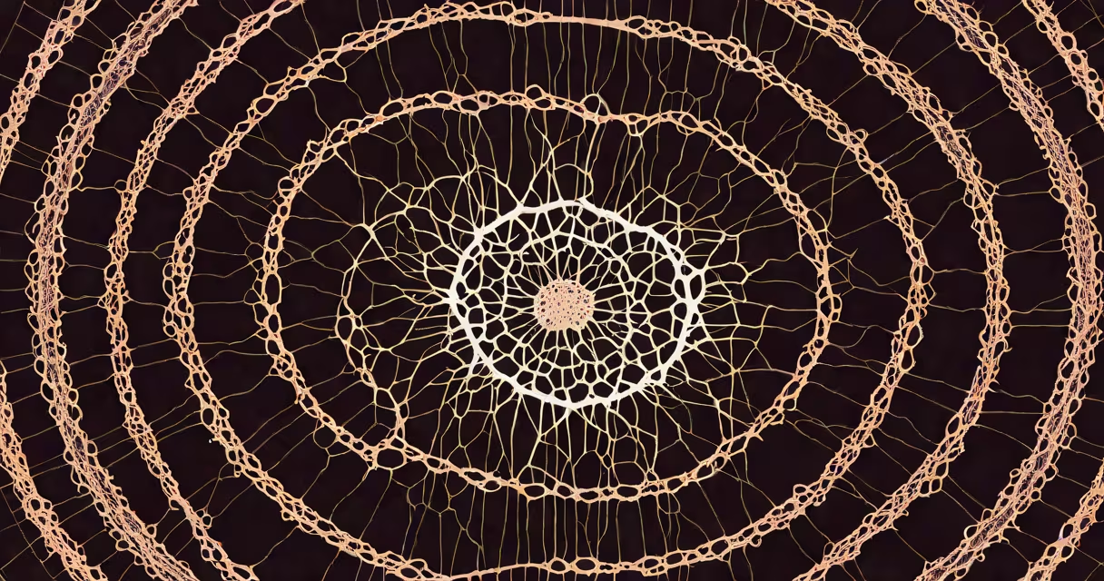
A radial scar is a benign breast lesion characterized by a central fibrous core with radiating duct-like structures that resemble spokes on a wheel. It appears on mammograms as a cluster of microcalcifications extending outward from a central point, often mimicking cancer. Though rare, it requires careful evaluation because of its potential to mimic early breast cancer.
Radial scars are not tumors. They are structural changes in breast tissue resulting from abnormal duct growth and fibrosis. They occur most frequently in women aged 40 to 60, though they can appear at any age. The term "radial" describes the star-shaped pattern seen on imaging, while "scar" reflects the fibrous tissue that forms the core.
According to the American College of Radiology (ACR), radial scars account for approximately 1-2% of all breast biopsies. While benign, they are associated with a slightly increased risk of developing breast cancer later in life-about 1.5 times higher than average. This makes follow-up essential, even after a benign diagnosis.
Radial scars develop when ducts in the breast grow abnormally and form a central core of fibrous tissue. Over time, these ducts branch out like rays, creating a starburst pattern. The process may result from prior injury, inflammation, or hormonal changes, though the exact cause remains unclear.
On mammography, radial scars typically show up as irregular clusters of microcalcifications. These calcifications are small (usually less than 1 mm) and arranged in a radial fashion. On ultrasound, they may appear as a hypoechoic mass with irregular borders, but they lack the solid structure of a tumor. Magnetic resonance imaging (MRI) can further clarify the architecture, showing a central area of low signal intensity surrounded by branching enhancement.
A 2021 study published in Radiology found that 78% of radial scars identified on mammography were confirmed histologically as benign. However, 22% showed atypical ductal hyperplasia (ADH) or low-grade ductal carcinoma in situ (DCIS)-conditions that require monitoring due to their link to future cancer development.
Pathologists classify radial scars based on their architectural features. The most common type is the classic radial scar, which shows a central fibrous nodule with multiple radiating ducts. Less common variants include the "atypical" radial scar, which displays cellular abnormalities in the duct lining and raises concern for precancerous changes.
| Type | Appearance on Imaging | Histological Features | Risk Level |
|---|---|---|---|
| Classic radial scar | Spoke-like calcifications, central density | Fibrous core, normal duct epithelium | Low (benign) |
| Atypical radial scar | Irregular calcifications, ill-defined margins | Ductal hyperplasia, nuclear atypia | Moderate (requires close follow-up) |
| Radial scar with DCIS | Clustered microcalcifications, rapid enhancement on MRI | Abnormal cells in ducts, confined to ducts | High (treatment recommended) |
Not all radial scars look the same. Some appear as dense masses on mammograms, others as subtle calcification patterns. The ACR's BI-RADS® classification system assigns radial scars to category 4 or 5 depending on their appearance and clinical context. A category 4 finding suggests a 2% to 95% chance of malignancy, requiring biopsy.
Radial scars are often mistaken for invasive ductal carcinoma due to their irregular shape and calcification patterns. In one retrospective review of 1,200 breast biopsies, 13% of radial scars were initially misclassified as suspicious for cancer. This highlights the challenge of distinguishing them from early-stage tumors.
The key difference lies in tissue architecture. Cancer cells invade surrounding tissue, while radial scars remain contained within ducts and fibrous stroma. Histopathology confirms this: under a microscope, cancer shows infiltrative growth, whereas radial scars display orderly, branching ducts with intact basement membranes.
I once reviewed a case where a 52-year-old woman had a radial scar detected on a routine screening mammogram. The calcifications were clustered and irregular. Initial radiologist interpretation flagged it as suspicious. After a core needle biopsy, pathology revealed a classic radial scar with no atypia. The patient was reassured and placed on annual surveillance. This case illustrates how imaging alone cannot confirm the diagnosis-tissue analysis is essential.
When a radial scar is found on imaging, the standard approach is surgical excision or core biopsy with pathological confirmation. The American Society of Breast Surgeons recommends complete removal of the lesion if it is visible on imaging and cannot be ruled out as malignant.
If the biopsy shows only benign tissue with no atypia, follow-up with annual screening mammograms is sufficient. However, if atypical ductal hyperplasia (ADH) or DCIS is found, the recommendation shifts to more frequent imaging-every 6 months-and consideration of risk-reducing strategies such as tamoxifen or genetic counseling.
For patients with a history of radial scar and ADH, the lifetime risk of developing breast cancer increases to about 8%. This is higher than the general population risk of 12%. The National Complete Cancer Network (NCCN) guidelines recommend discussing enhanced screening options, including annual breast MRI, for high-risk individuals.
Many patients assume that any lesion labeled "scar" is harmless. But radial scars are not the same as post-surgical scars. Post-surgical scars are linear, well-defined, and often located near previous incisions. Radial scars are diffuse, centered on ducts, and unrelated to trauma.
Another misconception is that radial scars always require surgery. Not true. If the lesion is small, well-circumscribed, and the biopsy results are benign, active surveillance may be appropriate. The decision depends on size, location, and patient risk factors.
Finally, some believe that radial scars are linked to breast implants or hormone replacement therapy. There is no evidence supporting this. Studies have shown no correlation between radial scar incidence and hormone use or implant presence.
Understanding what a radial scar is helps reduce anxiety and supports informed decisions. It is a benign finding in most cases, but its resemblance to cancer demands accurate diagnosis through biopsy and pathology. Early detection and proper management prevent unnecessary treatment while ensuring long-term safety.
For more on other lung-related findings, see here.