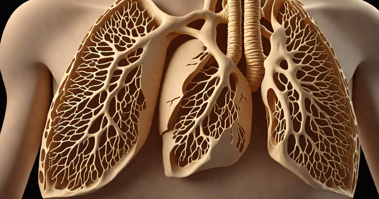
A scar on the lungs visible on a chest CT or X-ray indicates healed tissue damage from a prior injury, infection, or inflammatory condition. The scar is typically stable and does not grow, but it may affect lung function if extensive.
Scars form when lung tissue repairs itself after damage. The body replaces injured alveoli and connective tissue with dense, fibrous material. This process is called fibrosis. Scars appear as white or gray areas on imaging scans due to their higher density compared to healthy lung tissue.
Most scars result from resolved pneumonia, tuberculosis, or exposure to irritants like smoke or chemicals. They do not disappear and remain visible on follow-up scans. While they don't always cause symptoms, large or widespread scars can reduce oxygen exchange capacity.
Scars show up on high-resolution CT scans, which detect subtle differences in tissue density. On a CT, normal lung tissue appears dark because it contains air. Scarred areas appear lighter (hyperdense) due to the replacement of air spaces with fibrous tissue.
Radiologists describe lung scarring using specific patterns. Linear or reticular opacities along the lung bases suggest chronic changes. Honeycombing-small, cyst-like spaces surrounded by thick walls-is a sign of advanced fibrosis. These patterns help distinguish between old scarring and active disease.
For example, a 65-year-old patient with a history of smoking and recurrent bronchitis had a routine CT scan. The scan revealed bilateral basal linear opacities and mild volume loss. A follow-up six months later showed no change. The radiologist concluded the findings were consistent with stable, healed scarring from past respiratory infections.
Lung scarring varies in appearance and origin. The most common types include:
| Type | Appearance on Imaging | Common Causes |
|---|---|---|
| Linear fibrosis | Thin, straight lines extending from pleura inward | Pneumonia, post-surgical healing, radiation |
| Reticular pattern | Network of fine lines, often in lower lobes | Tuberculosis, hypersensitivity pneumonitis, occupational dust exposure |
| Honeycombing | Clustered small cysts with thick walls | Pulmonary fibrosis, long-term interstitial lung disease |
Not all scarring results from illness. Some people develop scars after surgery or trauma. Others have genetic predispositions to fibrosis, such as in idiopathic pulmonary fibrosis (IPF), where scarring occurs without a known trigger. IPF affects about 14 in 100,000 adults in the U.S., according to the American Lung Association.
One key distinction: scarring does not mean active disease. Unlike acute pneumonia, which shows fluid and consolidation, scars are static. They don't spread or worsen unless new injury occurs. This stability is critical for diagnosis and management.
Most lung scars do not cause symptoms and require no treatment. However, if scarring leads to reduced lung volume or impaired gas exchange, patients may experience shortness of breath during exertion.
Doctors assess scar impact through pulmonary function tests (PFTs). These measure how much air the lungs can hold and how efficiently oxygen moves into the blood. A patient with diffuse scarring may show reduced diffusing capacity (DLCO), indicating poor oxygen transfer.
For example, a 58-year-old woman with a history of rheumatoid arthritis developed fatigue and mild dyspnea. Her CT showed bilateral upper lobe reticular opacities. PFTs confirmed a 30% reduction in DLCO. She was referred to a pulmonologist, who diagnosed early-stage interstitial lung disease linked to her autoimmune condition. Treatment focused on managing inflammation to prevent further scarring.
Many assume a lung scar means cancer or ongoing disease. This is incorrect. Scars are not malignant. They do not grow or metastasize. A biopsy is rarely needed unless the lesion has atypical features like nodularity or growth over time.
Another misconception is that scars always impair breathing. In reality, small or localized scars rarely affect function. Only when scarring covers more than 30% of lung volume does it reduce respiratory reserve. The Global Initiative for Chronic Obstructive Lung Disease (GOLD) guidelines state that functional decline correlates with extent, not presence, of scarring.
One non-obvious fact: some scars can mimic active disease on imaging. For instance, a fibrotic scar may resemble a tumor if it has irregular borders. Radiologists use temporal comparison-looking at prior scans-to confirm stability. If a lesion hasn't changed size or shape over two years, it's almost certainly scar tissue.
If a scar is detected incidentally, doctors typically recommend monitoring rather than intervention. Follow-up imaging is advised only if symptoms develop or if new abnormalities appear.
Patients should review their medical history for past infections, exposures, or autoimmune conditions. Smoking history matters-smokers are more likely to develop fibrotic changes even without active disease. Quitting smoking reduces risk of progression.
A scar on the lungs is a record of past damage. It's not a current threat. Most people live normally with stable scars. The focus shifts from treating the scar to managing underlying causes and preventing further injury.