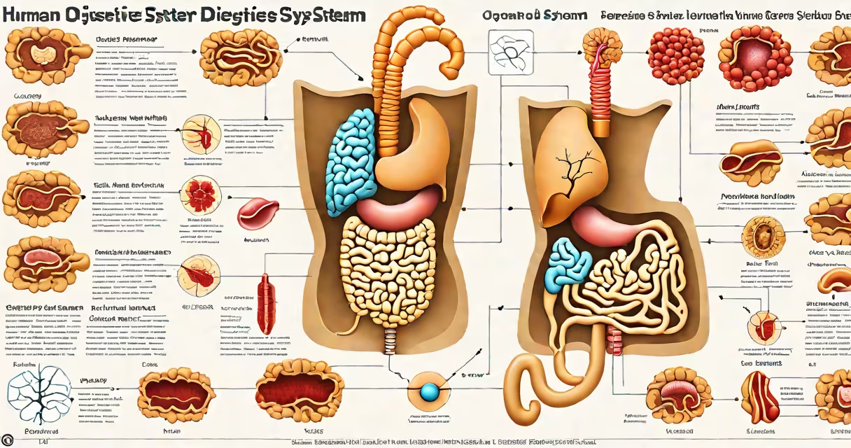
A human digestive system diagram labeled organs is a visual representation that maps the pathway of food through the gastrointestinal tract, identifying each organ involved in breaking down food, absorbing nutrients, and eliminating waste. These diagrams show the sequence from the mouth to the anus, highlighting structural relationships and functional roles.
Such diagrams are essential tools in biology, medicine, and health education. They display the esophagus, stomach, small intestine (duodenum, jejunum, ileum), large intestine (colon), rectum, and anus, along with accessory organs like the liver, gallbladder, and pancreas. Each structure is labeled with its name and function, enabling quick reference and learning.
Medical textbooks, classroom materials, and patient education resources commonly use these diagrams. They are designed to reflect anatomical accuracy based on cadaver studies and clinical imaging. The National Institutes of Health (NIH) and the American Association of Anatomists both endorse standardized labeling systems for consistency across educational content.
The digestive process begins in the mouth, where chewing and saliva initiate carbohydrate breakdown. Food travels down the esophagus via peristalsis-wave-like muscle contractions-and enters the stomach. Here, gastric juices containing hydrochloric acid and pepsin break proteins into smaller peptides.
In the small intestine, most nutrient absorption occurs. The duodenum receives bile from the liver (stored in the gallbladder) and pancreatic enzymes (from the pancreas), which further digest fats, carbohydrates, and proteins. The inner lining of the small intestine features villi and microvilli, increasing surface area for efficient absorption.
The remaining material moves into the large intestine, where water and electrolytes are reclaimed. Bacteria in the colon ferment undigested fiber, producing gases and certain vitamins like K and B12. Waste forms into stool and exits through the rectum and anus.
Each organ in the human digestive system plays a distinct role. The following table outlines the primary functions of major components:
| Organ | Primary Function | Key Output |
|---|---|---|
| Mouth | Mechanical breakdown via chewing; initial enzymatic digestion via salivary amylase | Chyme (partially digested food mass) |
| Stomach | Protein digestion using pepsin; acid sterilization of ingested microbes | Chyme with reduced pH (1.5-3.5) |
| Small Intestine | Most nutrient absorption; enzyme action from brush border and pancreas | Glucose, amino acids, fatty acids absorbed into bloodstream |
| Large Intestine | Water reabsorption; bacterial fermentation of fiber | Stool formation; vitamin K synthesis |
| Liver | Bile production; detoxification of blood; protein synthesis | Bile stored in gallbladder |
| Pancreas | Secretion of bicarbonate and digestive enzymes (lipase, protease, amylase) | Enzymes released into duodenum |
One non-obvious fact: the small intestine is approximately 6 meters long in adults-longer than a standard basketball court. Despite its name, it's the longest segment of the digestive tract and responsible for absorbing about 90% of dietary nutrients.
Many people assume the large intestine is primarily responsible for digestion. In reality, it handles water recovery and waste compaction. Digestion-chemical breakdown of food-occurs mainly in the small intestine. The large intestine's role is more about processing leftovers.
Another misconception is that the gallbladder produces bile. It does not. The liver synthesizes bile, which is stored and concentrated in the gallbladder before release into the duodenum during fat-rich meals. Gallstones form when cholesterol or bilirubin precipitates in the gallbladder, blocking bile flow.
Some diagrams incorrectly place the appendix near the stomach or suggest it aids digestion. The appendix is a vestigial organ with no known digestive function. It may play a minor role in immune cell development in the gut-associated lymphoid tissue (GALT), but its removal doesn't impair digestion.
Health professionals use labeled digestive system diagrams to explain conditions like gastroesophageal reflux disease (GERD), Crohn's disease, and irritable bowel syndrome (IBS). For example, a patient with chronic heartburn may benefit from seeing how the lower esophageal sphincter fails to close properly, allowing acid to reflux into the esophagus.
In a clinical setting, a surgeon preparing for laparoscopic surgery reviews a labeled diagram to identify anatomical landmarks. During a cholecystectomy (gallbladder removal), the cystic duct and artery must be precisely located to avoid injury to the common bile duct-a complication that occurs in roughly 0.5% of cases, according to the American College of Surgeons.
I once observed a medical student struggle to locate the duodenum during a dissection. A labeled diagram clarified that the duodenum curves around the head of the pancreas-the "C-shaped" loop visible in cross-section. That single visual cue resolved confusion and accelerated learning.
The digestive system operates in tandem with other bodily systems. The nervous system regulates motility through the enteric nervous system-the "second brain" of the gut. Hormones like ghrelin and leptin control hunger and satiety, linking digestion to energy balance.
Always consult a validated source when interpreting a human digestive system diagram labeled organs. The Visible Body platform and the Visible Human Project by the NIH provide peer-reviewed, high-resolution models used in academic institutions worldwide.
These diagrams are not just educational-they're diagnostic tools. When a doctor sees a patient with unexplained weight loss, a labeled diagram helps determine whether the issue lies in malabsorption (small intestine), maldigestion (pancreas), or obstruction (colon).