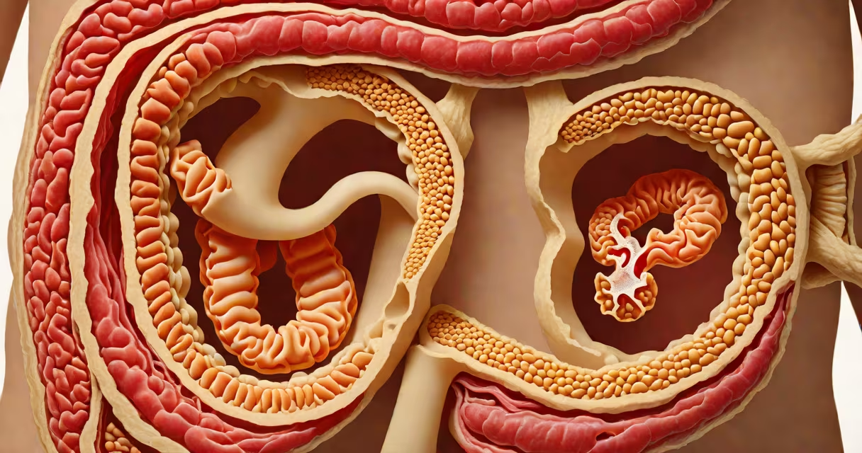
The cecum is the initial segment of the large intestine, situated in the lower right quadrant of the abdomen. It receives chyme from the ileum via the ileocecal valve and marks the beginning of the colon's process through the abdominal cavity.
Located just below the ileocecal sphincter, the cecum acts as a transitional chamber between the small and large intestines. It measures approximately 6-8 cm in length and 7-9 cm in diameter, forming a pouch-like structure that collects undigested material from the small intestine before passing it along the ascending colon.
Medical imaging confirms the cecum lies near the right iliac fossa, often palpable during physical exams in thin individuals. Its position is consistent across adults, though slight variation occurs due to bowel motility and individual anatomy.
The cecum plays a key role in water absorption and fermentation of residual carbohydrates. As chyme enters from the ileum, the cecum begins the process of transforming liquid waste into solid stool by extracting remaining water and electrolytes.
Bacterial fermentation in the cecum produces short-chain fatty acids such as butyrate, which nourish colonic epithelial cells. This function supports gut health and contributes to immune regulation in the lower gastrointestinal tract.
A 2021 study published in Gastroenterology found that patients with reduced cecal motility had higher rates of constipation and altered microbiome composition, underscoring the organ's functional significance beyond mere transit.
The cecum sits at the junction of the small and large intestines. It connects to the ascending colon via the cecocolic flexure and is anchored by the mesentery of the small intestine and the mesocolon.
Several structures lie in close proximity:
During surgical procedures, the cecum serves as a landmark for identifying the start of the colon. Surgeons use its position to verify correct anatomical orientation during colectomies and laparoscopic interventions.
Many assume the cecum is the site of appendicitis onset. While the appendix arises from the cecum, inflammation typically begins within the appendix lumen due to obstruction, not in the cecum itself. The cecum may become inflamed secondarily if the appendix ruptures.
Another misconception is that the cecum is always visible on standard abdominal X-rays. In reality, it appears only when distended with gas or fecal matter. A normal KUB (kidney-ureter-bladder) X-ray rarely shows the cecum unless it contains significant air.
When diagnosing right lower quadrant pain, clinicians differentiate cecal involvement from other causes. For example, cecal volvulus-a rare twisting of the cecum-can mimic appendicitis but requires surgical correction. CT scans show a "coffee bean" sign in such cases, distinguishing them from typical appendicitis.
In a 2019 case report from the Journal of Emergency Medicine, a 68-year-old man presented with acute abdominal pain localized to the right lower quadrant. Initial ultrasound showed no evidence of appendicitis. A subsequent CT scan revealed a dilated cecum with a twisted mesentery, confirming cecal volvulus.
Imaging displayed a characteristic "whirlpool" sign around the cecal base and a twisted vascular pedicle. The patient underwent emergency surgery to detorse the cecum and resect the redundant mesentery. Postoperative recovery was uneventful, highlighting the importance of accurate anatomical localization.
Understanding the cecum's role requires distinguishing it from adjacent colon regions. The following table outlines key differences:
| Feature | Cecum | Ascending Colon | Transverse Colon |
|---|---|---|---|
| Length | 6-8 cm | 15-20 cm | 30-40 cm |
| Function | Receives chyme, initiates water absorption | Continues water and electrolyte absorption | Final water absorption and stool formation |
| Position | Lower right abdomen | Right side, ascends toward liver | Across upper abdomen, from right to left |
| Pathology Risk | Volvulus, rupture | Diverticulosis, cancer | Cancer, ischemia |
Each segment has distinct clinical risks. The cecum's limited mobility increases risk of volvulus, while the transverse colon is more prone to ischemia due to its long vascular supply.
During a routine laparoscopic appendectomy, I observed the cecum's position firsthand. After insufflating the abdomen, the cecum appeared as a rounded, pale structure beneath the right lower quadrant. The appendix extended from its posterior wall, and the ileocecal valve was visible as a fold of mucosa.
Using a grasper, I gently lifted the cecum to expose the appendix base. The consistency of the tissue and the presence of fat pads confirmed normal anatomy. This direct view reinforced how central the cecum is to both diagnostic and surgical planning.
The cecum is the entry point of the large intestine, located in the lower right abdomen. It receives contents from the small intestine and initiates the colon's role in fluid conservation and microbial processing. Its position is critical for diagnosing conditions like appendicitis and cecal volvulus.