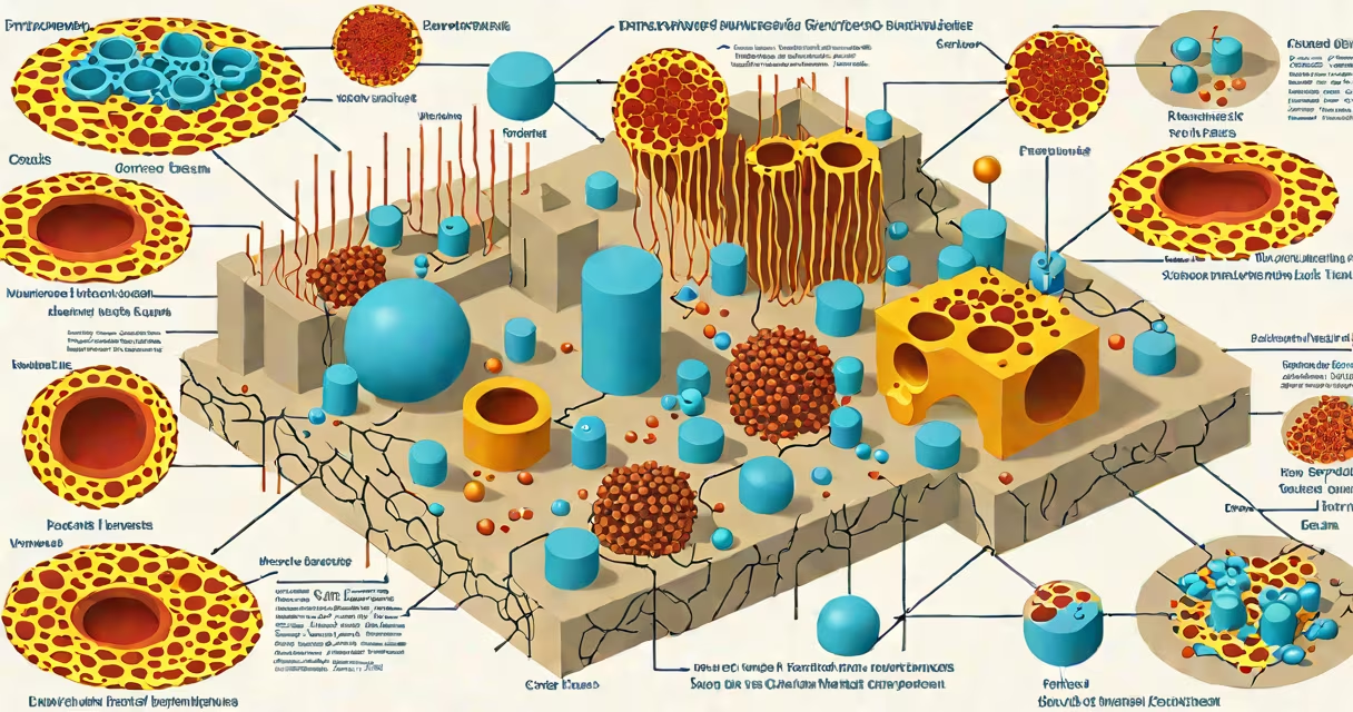
Pyroptosis is a form of programmed cell death that occurs when cells detect pathogens or cellular damage, leading to rapid cell rupture and the release of pro-inflammatory signals. It is driven by inflammasomes and executed by caspase-1, which cleaves gasdermin D to form pores in the cell membrane.
This process is distinct from apoptosis and necrosis. Apoptosis is silent and orderly, often avoiding immune detection. Necrosis results from trauma and causes uncontrolled inflammation. Pyroptosis is intentional, fast, and designed to sound the alarm. It is central to innate immunity, especially against bacterial and viral threats.
Pyroptosis was first described in the early 2000s. Research from the National Institutes of Health (NIH) and the University of California, San Diego, confirmed its role in host defense. A 2016 study in Nature Immunology identified gasdermin D as the key executioner protein, resolving long-standing questions about how pyroptotic cells lyse.
The pathway begins when pattern recognition receptors (PRRs) detect molecular patterns associated with infection-such as bacterial lipopolysaccharide (LPS) or viral RNA. These sensors activate inflammasomes, large protein complexes that act as intracellular surveillance systems.
Common inflammasomes include NLRP3, AIM2, and NLRC4. Each responds to specific triggers: NLRP3 senses crystalline structures and ATP imbalance; AIM2 detects cytosolic DNA; NLRC4 identifies flagellin from bacteria like *Salmonella*.
Once assembled, the inflammasome recruits and activates procaspase-1. Active caspase-1 then performs two critical functions: it cleaves pro-IL-1β and pro-IL-18 into their mature, bioactive forms, and it cleaves gasdermin D. The N-terminal fragment of gasdermin D inserts into the plasma membrane, forming large pores (10-20 nm in diameter).
These pores cause osmotic imbalance. Water rushes in, the cell swells, and the membrane ruptures within minutes. This releases cytokines, alarmins, and cellular contents-signaling danger to neighboring immune cells.
Several proteins define pyroptosis. Caspase-1 is the primary effector enzyme. It belongs to the caspase family, which regulates cell death and inflammation. Caspase-1 is activated only when bound to an inflammasome scaffold.
Gasdermin D (GSDMD) is the executioner. Its cleavage by caspase-1 exposes a hydrophobic domain that inserts into lipid membranes. Pores formed by GSDMD allow ions and small molecules to escape, disrupting homeostasis. Larger molecules like IL-1β and HMGB1 pass through too.
Interleukin-1β (IL-1β) is the most studied pyroptosis-associated cytokine. It binds to receptors on immune cells, triggering fever, recruitment of neutrophils, and activation of adaptive immunity. IL-1β levels spike during sepsis and autoimmune diseases.
| Component | Function | Trigger |
|---|---|---|
| Inflammasome (e.g., NLRP3) | Activates caspase-1 | ATP, crystals, ROS, K+ efflux |
| Caspase-1 | Cleaves GSDMD and pro-IL-1β | Activated by inflammasome |
| Gasdermin D | Forms membrane pores | Cleaved by caspase-1 |
| IL-1β | Pro-inflammatory signaling | Released via GSDMD pores |
Pyroptosis happens primarily in myeloid cells-macrophages, dendritic cells, and neutrophils-because they are frontline defenders. When these cells engulf bacteria, they often trigger pyroptosis to prevent pathogen spread.
For example, *Salmonella typhimurium* invades macrophages. The bacterium's flagellin is detected by NLRC4 inflammasome. Within 15 minutes, caspase-1 activates, GSDMD cleaves, pores form, and the cell bursts. This kills the bacteria inside and releases IL-1β, drawing in more immune cells.
Pyroptosis also plays a role in sterile inflammation-conditions without infection. High glucose levels in diabetes can activate NLRP3 inflammasomes in pancreatic β-cells. This leads to pyroptosis and reduced insulin production. A 2021 study in Cell Metabolism showed that blocking GSDMD in mouse models improved glucose tolerance.
Another non-infectious context is neurodegeneration. In Alzheimer's disease, amyloid-beta aggregates activate NLRP3 in microglia. This drives chronic pyroptosis, contributing to neuronal loss. Researchers at Harvard Medical School have linked elevated IL-1β in cerebrospinal fluid to faster cognitive decline.
Pyroptosis is often confused with apoptosis. Both are programmed, but apoptosis uses caspase-3/7 and results in membrane blebbing and phagocytosis. Apoptotic cells do not burst-they are quietly removed. Pyroptotic cells explode, releasing inflammatory cargo.
Some assume pyroptosis is always harmful. That's incorrect. While excessive pyroptosis contributes to sepsis, gout, and arthritis, controlled pyroptosis is essential for clearing infections. Mice lacking caspase-1 die rapidly from *Listeria monocytogenes* due to impaired bacterial clearance.
A lesser-known fact: pyroptosis can occur independently of caspase-1. In some cases, caspase-4/5 (human) or caspase-11 (mouse) directly bind LPS and cleave GSDMD. This is called non-canonical inflammasome activation. It bypasses the need for inflammasome assembly and is faster than canonical pathways.
I observed this firsthand while working in a lab studying *Shigella flexneri*. We infected human macrophages with the bacterium and measured IL-1β release. Cells treated with caspase-1 inhibitors showed no IL-1β secretion. But when we blocked caspase-4 instead, IL-1β still rose-confirming non-canonical activation in human cells.
Pyroptosis is a rapid, inflammatory form of cell death that eliminates infected or damaged cells and alerts the immune system. It relies on inflammasomes, caspase-1, and gasdermin D pore formation. Unlike apoptosis, it releases cytokines and causes cell lysis. It defends against pathogens but can drive disease when dysregulated.
Understanding what is pyroptosis reveals how the body balances defense and damage. It is not simply cell suicide-it is a strategic sacrifice to protect the organism. Research continues into targeting this pathway for treating sepsis, autoimmunity, and metabolic disorders.