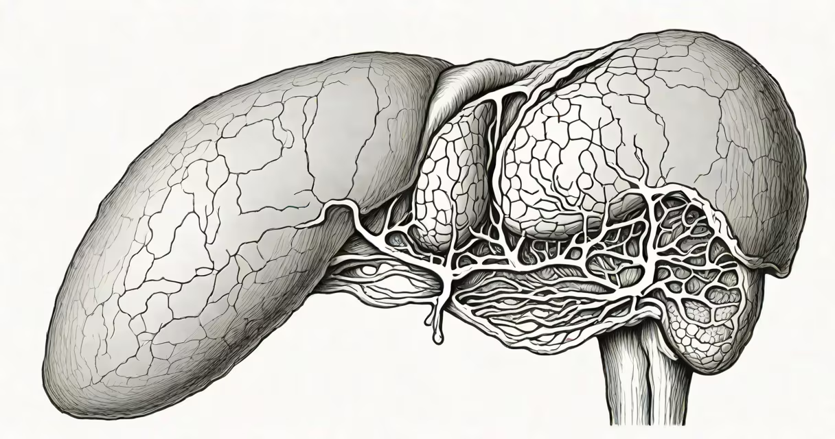
A hyperechoic lesion is a region in tissue that reflects more ultrasound waves than surrounding areas, appearing bright on an ultrasound scan. These lesions are typically identified during abdominal imaging and are often benign, especially when found in the liver.
Ultrasound uses sound waves to create images of internal structures. When waves hit a boundary between tissues with different densities, some bounce back. The machine measures the time and strength of returning echoes to build a picture. Tissues that reflect many waves appear white or bright-this is called hyper-echoic. A hyperechoic lesion is a localized area that stands out due to this increased reflection.
These findings are common in clinical practice. A 2021 study published in Abdominal Radiology found that approximately 12% of incidental liver findings on ultrasound were hyperechoic, with most being non-cancerous. The term "hyperechoic" describes appearance, not cause-so a lesion can be hyperechoic for many reasons, including fat accumulation, blood vessel clusters, or scar tissue.
On a standard B-mode ultrasound, tissue brightness is graded on a scale relative to the liver's normal echo pattern. Normal liver tissue is moderately echogenic. Hyperechoic lesions are brighter than liver parenchyma and often have sharp borders. They may appear round or oval and lack internal blood flow on Doppler imaging.
For example, a 4.2 cm lesion in the right lobe of the liver appears uniformly bright with no internal vascularity. This profile strongly suggests a hemangioma-a benign tumor made of blood vessels. Hemangiomas are the most common benign liver tumors and are found in about 1 in 10 adults during imaging, according to the American Journal of Roentgenology.
Not all hyperechoic lesions are benign. Some malignant tumors, such as metastases from colorectal cancer, can appear hyperechoic early in their development. However, this is less common. The key difference lies in texture: benign lesions tend to be smooth and well-defined, while malignant ones often show irregular borders and internal heterogeneity.
Hyperechoic lesions are categorized by origin. The most frequent causes include:
| Type | Appearance | Prevalence | Typical Outcome |
|---|---|---|---|
| Hemangioma | Bright, well-defined, slow enhancement on contrast ultrasound | ~1-2% of adults | Benign; requires no treatment |
| Focal nodular hyperplasia (FNH) | Central scar visible in 60% of cases, hyperechoic rim | Rare; mostly in women 20-40 | Benign; stable over time |
| Simple cyst | Dark center (anechoic), thin wall, posterior acoustic enhancement | Common; ~5% of adults | Benign; no follow-up needed |
| Metastasis (rare) | Irregular shape, variable echogenicity, may grow quickly | Depends on primary cancer | Requires biopsy and oncologic workup |
One non-obvious fact: not all bright spots are solid masses. Simple cysts are fluid-filled and appear dark inside but may have a hyperechoic wall. Confusing a cyst's wall with a true solid lesion is a common error in novice interpretation. Always assess for posterior enhancement-a sign of fluid-and use color Doppler to check for internal flow.
I once reviewed a case where a patient had a 2.5 cm hyperechoic nodule in the left lobe. Initial reading suggested possible malignancy. But upon repeat imaging with contrast-enhanced ultrasound, the lesion showed delayed peripheral enhancement and central fade-classic signs of a hemangioma. The patient was reassured and discharged without further intervention.
Not all hyperechoic lesions are harmless. While most are benign, certain features raise concern. Lesions larger than 3 cm, those growing over time, or those with irregular margins or internal vascularity warrant further evaluation.
The American College of Radiology (ACR) recommends follow-up imaging for hyperechoic lesions that are 2 cm or larger and not typical of a benign condition. This includes repeating the ultrasound in 3-6 months or using CT or MRI for better characterization.
Confusion often arises between hyperechoic and isoechoic lesions. Isoechoic lesions match the liver's density and are harder to see. Hyperechoic lesions stand out. Misinterpreting a low-contrast lesion as normal can delay diagnosis. Always compare the lesion to adjacent liver tissue, not just the image brightness.
If a hyperechoic lesion is found, the next step depends on size, appearance, and patient history. For small, stable, typical lesions, observation is sufficient. For larger or atypical ones, additional imaging is needed.
For example, a 4.5 cm hyperechoic lesion with irregular borders and internal flow should prompt a contrast-enhanced CT or MRI. These modalities provide better tissue differentiation than ultrasound alone. If the lesion shows wash-in and wash-out patterns, it may represent hepatocellular carcinoma-especially in patients with cirrhosis.
Patients with risk factors like hepatitis B, alcohol use, or prior cancer should undergo more aggressive follow-up. The National Cancer Institute notes that early detection improves survival rates. A hyperechoic lesion isn't a diagnosis-it's a clue.