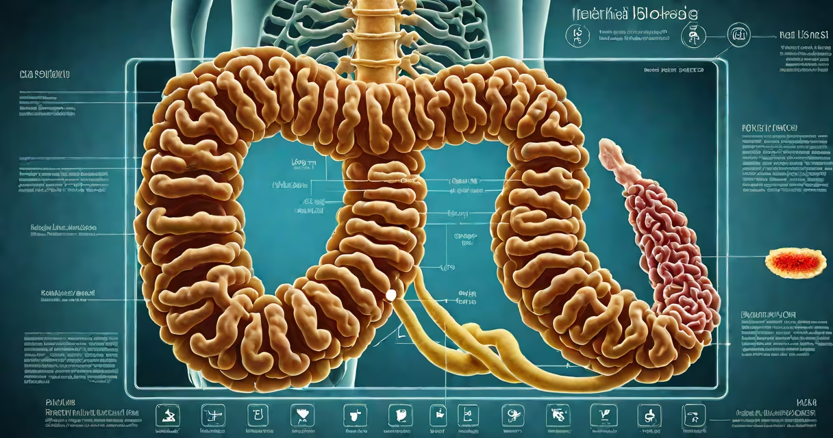
Intestinal blockage is diagnosed through a combination of patient history, physical examination, blood tests, and imaging scans-most commonly abdominal X-rays, CT scans, and ultrasound-to identify obstructions in the small or large intestine.
Doctors begin by assessing symptoms such as severe abdominal pain, bloating, vomiting, and inability to pass stool or gas. These signs suggest a partial or complete blockage. A physical exam may reveal tenderness, distension, or absent bowel sounds. The presence of fever or dehydration raises concern for complications like ischemia or perforation.
Imaging is essential. An upright abdominal X-ray shows air-fluid levels and dilated loops of bowel-hallmarks of obstruction. A CT scan with oral contrast provides higher accuracy, identifying the location, cause (e.g., tumor, hernia, adhesions), and severity. Ultrasound is less common but useful in children or pregnant patients to avoid radiation.
Diagnosis follows a tiered approach. Initial assessment includes important signs, symptom timeline, past surgeries, and medication use. Patients with prior abdominal surgery have a higher risk of adhesive blockages.
Blood tests measure electrolyte imbalances, kidney function, and signs of infection. Elevated white blood cell count or lactate levels indicate possible bowel ischemia-a surgical emergency. Dehydration markers like high BUN-to-creatinine ratio support fluid loss from vomiting.
Imaging confirms the diagnosis. A single abdominal X-ray detects obstruction in about 80% of cases. A CT scan improves detection to over 95%, especially when contrast is used. For example, a 2021 study in *Radiology* found CT had 96% sensitivity and 94% specificity for diagnosing mechanical bowel obstruction.
| Test | Use Case | Accuracy |
|---|---|---|
| Abdominal X-ray (upright) | First-line screening | ~80% |
| CT scan with contrast | Definitive diagnosis, localization | 94-96% |
| Ultrasound | Pregnant patients, pediatric cases | ~75% (operator-dependent) |
One non-obvious point: a normal X-ray doesn't rule out obstruction. In early or partial blockages, changes may not yet appear. A negative X-ray with persistent symptoms requires further imaging. CT remains the gold standard when uncertainty exists.
Blockages stem from mechanical causes-tumors, hernias, volvulus, impacted feces-or functional issues like paralytic ileus. Adhesions from prior surgery account for 60% of small bowel obstructions, according to the American Society of Colon and Rectal Surgeons.
Paralytic ileus mimics mechanical obstruction but lacks visible blockage on imaging. It results from medications, infections, or metabolic disturbances. Misdiagnosis occurs when doctors treat symptoms without confirming obstruction. A 2019 review in *The New England Journal of Medicine* noted that up to 15% of suspected obstructions are actually functional ileus.
Patients with chronic constipation may present with symptoms similar to blockage. A misdiagnosis can delay treatment for underlying conditions like Hirschsprung's disease or colonic inertia. Always consider patient history: recent travel, dietary changes, or medication use (e.g., opioids) can influence presentation.
A 68-year-old man presented with six hours of crampy abdominal pain, vomiting bile, and no bowel movement. He had a history of colon cancer resection five years prior. On exam, his abdomen was distended with hyperactive bowel sounds.
An upright abdominal X-ray showed multiple air-fluid levels and dilated small bowel loops. A CT scan confirmed a partial obstruction at the jejunum with thickened walls and mesenteric stranding-indicating inflammation. The cause was a recurrent tumor. Surgery was scheduled within 24 hours.
This case illustrates the diagnostic cascade: symptoms → physical exam → X-ray → CT confirmation → intervention. Without imaging, the tumor might have progressed to complete obstruction or perforation.
Delays happen when patients delay seeking care or when providers overlook subtle signs. A 2020 study in *Annals of Surgery* found that 30% of patients with obstructive symptoms waited over 24 hours before presenting to emergency departments. This delay increases the risk of bowel necrosis.
Another error is relying solely on lab results. Normal electrolytes don't exclude obstruction. One patient with a small bowel obstruction had normal sodium and potassium levels but developed sepsis due to delayed diagnosis. Imaging remains critical even with stable labs.
I once observed a patient in a rural ER whose initial X-ray was interpreted as "normal." After worsening pain and fever, a repeat CT revealed a closed-loop obstruction requiring urgent surgery. The lesson: no test is infallible, and clinical judgment must evolve with new symptoms.